


Trauma: A direct blow to the bone, such as from a fall or car accident, can cause a fracture.
Osteoporosis: This condition causes the bones to weaken and become more fragile, increasing the risk of fractures.
Bone cancer: Malignant tumours can weaken the bones and make them more susceptible to fractures.
Metabolic disorders: Conditions like osteomalacia (softening of the bones) and Paget’s disease (abnormal bone growth) can make the bones more prone to fractures.
Genetic disorders: Some disorders, such as osteogenesis imperfecta (brittle bone disease), can make bones more fragile and susceptible to fractures.
A combination of physical examination and imaging tests is usually used to diagnose bone fractures.The most common imaging tests used to diagnose bone fractures are X-rays, computed tomography (CT) scans, and magnetic resonance imaging (MRI) scans.
During a physical examination, the doctor will feel tenderness, swelling, and deformity around the injured area. The doctor may also ask the person to move the injured limb in certain ways to see if the range of motion is limited or if there is any pain.
Imaging tests are often needed to confirm the diagnosis of a bone fracture and to determine the type and extent of the injury. X-rays are the most common type of imaging test used to diagnose bone fractures. They can show whether a bone is broken and sometimes reveal the type of break.
CT scans and MRI scans are more sensitive than X-rays and can show small fractures that may not be visible on an X-ray. CT scans and MRI scans can also show the surrounding soft tissue, which can be helpful in planning treatment.
Immobilization: This involves keeping the broken bone from moving by using a cast, splint, or brace. This is often the best option for fractures that are not displaced (the ends of the bone are still in alignment) and for those that are not open (the skin is not broken).
Surgery: Surgical intervention may be necessary to align the ends of the bone or to insert metal rods, pins, or screws to hold the bone in place. Surgery is also sometimes used to treat open fractures (those with a break in the skin).
Traction: This is a method of gradually pulling the bone ends into alignment using weights and pulleys. It is often used for fractures of the long bones in the arm or leg.
External fixation: This involves attaching metal screws, pins, or rods to the bone on the outside of the body. The metal devices are then connected to a stabilising frame that is worn on the outside of the body.
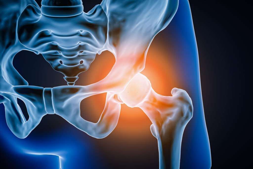
Internal fixation: This is similar to external fixation, but the metal screws, pins, or rods are placed inside the body, and the stabilising frame is placed on the outside. Internal fixation is often used for fractures of the pelvis or hip.
© 2024 Deepa Hospital. All Rights Reserved.